Endodontics: June 2011

Thursday 30 June 2011

Profile System - A new Niti Instruments


Profile System

Radial- Landed Flute & U-File Design
a  Shaping Coronal Portion of Canal
a  Manipulate in Difficult Access Area
a  60 degree Bullet-Nosed Tip
a  Cutting Angle 90 degree
a  Planes the Wall without Gouging & Self Threading
a  Even Stress Distribution

Benefits :-
J Improves Flexibility & Cutting  ,efficiency in Tighter or more Curved Canal.
J Fewer Files needed.
J Reduces Torsional Load , File Fatigue                 and Potential Separation.
J Reduces Contact Area between File and Dentine.


Sunday 26 June 2011

Ni Ti Instruments in endodontics


Ni Ti Instruments in Endodontics
Advantages :-


          Reduced instrumentation time.
           Simplified RC preparation.

Disadvantages  of stainless steel files :-

          Increased canal blockage &         Instrument breakage
           Insufficient canal debridement


Stainless Steel Files: Shortcomings


L Increased Time of  Canal Preparation
L Made Obturation Less Predictable
L Canal Transportation due to Design
L Excessive Dentine Removal


Nickel Titanium Rotary Instruments
Genesis
           Nickel Titanium Naval                    Ordinance Lab. (NITINOL)
           Property - ‘Superelasticity’                          (1960)


Tapered Preparation                     
          Improved cleaning ability
          Enhanced apical control of instruments
          Less dependable on exact length determination
          More dependable apical resistance
          Greater confidence of cone fit
          0.02 Taper
           0.06 Taper

Wednesday 22 June 2011

Immediate Implant placement

Immediate Implant placement


RVG before implant placement with root piece with upper right central incisor .


Immediately after the placement of implant


Monday 20 June 2011

Avulsion

Extraction site after avulsion of deciduous teeth due to trauma
Trauma to four year old child leading to exfoliation of decicuous left central incisor along with the injury to right deciduous central incisor .This RVG shows that injury might have extended upto permenent left central inciosr tooth bud . 

Friday 17 June 2011

Todays case with root canal treatment

This is left mandibular first molar immediately after obturation with temporary restoration with zinc oxide eugenol cement with grade I furacation involvement.

Tuesday 14 June 2011

Monday 13 June 2011

Length of master cone determiantion

Length of master cone determiantion
Length of Master cone determiation is done by various methods .They are (1) Tug back method and (2) Radiographic method .

Sunday 12 June 2011

Postoturation restoration / Restoration after obturation

Postoturation  restoration


This particular radiograph shows postobturation restoration with mandibular first molar and second madibular molar shows errupting second molar with open apex .
This postobturation restoration shows restoration is silver amalgam which is distocclusal in type showing scattered silver amalgam particles and restoration is not well contoured distally with overhang seen cervically.

Saturday 11 June 2011

Obturation with tooth having internal resorption.

Obturation with tooth having internal resorption.

This is RVG / radiograph having trauma couple of years ago which have perapical abscess with draining sinus .Such a long standing perapical pathosis leads to internal resorption which after oturation will shape the canal like this RVG /radiograph.

Thursday 9 June 2011

Molar Single sitting root canal treatment

Molar Single Sitting Root Canal Treatment
This RVG / Radiograph shows Oturation of second mandibualr molar done in a single sitting under proper antibiotic and NSAIDs coverage in order to reduce the intensity of pain and edema .
This particualr patient do not have  edema but pain is present which is because of perapical pathosis .This pathosis will take some time to subside.
This sort of single sitting root canal treatment will reduce the patient's suffering more rapidly than multiple sitting root canal treatment which is traditionally followed.
If you are take into consideration all aseptic condition and use proper intracanl medicament to make canal absolutely clean and microoraganism free then the success as well as prognosis will be good in such cases.

Premolar Obturation

Premolar Obturation 


This is second premolar immedaitely after oturation 

Tuesday 7 June 2011

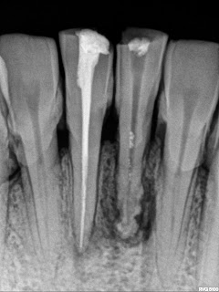
External root resorption with lower anterior teeth due to chronic perodontitis

External root resorption with lower anterior teeth due to chronic perodontitis
This radiograph shows generalised chronic periodontitis with horizontal bone loss with external root resorption at the apical third of the root with calculus attached cervically with all teeth / incisors seen in the radiograph. Both the central incisors are attrided incisally with less damage to the bone with root canal treated incisor and damage seen with other incisor is more.

Monday 6 June 2011

Fractured central incisor


Fracutred Central incisor in the middle third of the root due to trauma and best treatment plan for this tooth is wait and watch for few weeks for the improvement of mobility and then decide. Ideally the treatment for such type of fracture in the middle third of the root is extraction becasue prognosis is poor .